We have a highly interactive and well-funded group of investigators, both within the Division of Nephrology and throughout Washington University School of Medicine, who study kidney disease. Research collaborations include investigators from multiple disciplines, including cell biology, molecular biology, genomics, developmental biology and physiology, who all have a central aim to understand the mechanisms of renal disease and dysfunction.
Processes being studied include:
- Matrix deposition
- Trafficking
- Cell-cell interactions
- Ion transport
- Fate determination
- Pattern and boundary formation
- Cell migration, proliferation, renewal and death
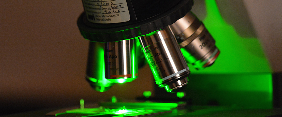
Newer technologies range from single cell genomics to 4D time lapse microscopy and stochastic optical reconstruction microscopy (STORM). Other technologies relevant to renal research include kidney injury models such as ischemia reperfusion, ureteral obstruction, 5/6 nephrectomy, parabiosis, toxic injury (cisplatin, folate, aristolochic acid), kidney, ureter or whole GU organ culture using time lapse microscopy, cilia and centrosome analysis, ureteral obstruction models, glomerular filtration analysis, ureter peristalsis and genomic analysis.