The Renal Biopsy Service, a new in-house program, is nearly ready to launch. Assistant Professor of Medicine Fadi Tohme, MD, who holds dual appointments in the Division of Nephrology and the Division of Anesthesiology, will be director of the service.

Dr. Fadi Tohme
The new program, in which our own nephrologists will perform ultrasound-guided kidney biopsies, will be a valuable resource in teaching this skill to incoming fellows.
The renal biopsy, most commonly performed when the cause of kidney damage is uncertain, is an indispensable tool in the diagnosis, prognosis and management of patients with renal diseases. However, only a handful of nephrology programs in the United States teach ultrasound-guided kidney biopsies to their fellows.
“We will certainly be on the nationwide forefront of programs that offer such a service to patients, while also educating the fellows,” says Dr. Tohme. “This is really something positive for our program.”
Making the kidney biopsy an integral part of nephrology fellow training has been in the planning since Dr. Tohme joined the division in July 2017.
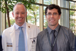
Dr. Benjamin Humphreys (left) and Dr. Andrew Malone, at last fall’s 5,000 Kidney Transplant celebration.
“Our division chief, Dr. Benjamin Humphreys, was very keen on the thought of us, the nephrologists, mastering this technique so that we can teach the fellows how to do it properly,” says Tohme, “so that that they can get that skill and be comfortable with it once they’ve graduated from the program.”
The plan was that it would take about a year to get the service up and running. “It has been a long process, but we will be able to launch it in September or October!”
The current procedure for obtaining a kidney biopsy here is the two-person method. A radiology technician uses ultrasound (or CT in some cases) to locate and image the kidney, and then a nephrologist obtains the biopsy using a spring-loaded needle. Under the new system, the nephrologist will be the one manipulating both the ultrasound and the biopsy needle. Tohme points out that, “This way, we are taking more responsibility, and it is more satisfying as well when we are able to do the whole procedure ourselves.”
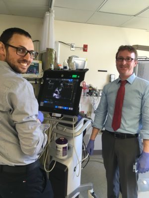
Dr. Tohme (left) and Dr. Frank O’Brien (right) testing out the new Nephrology ultrasound machine.
Our nephrologists have been conducting ultrasound-guided biopsies for the transplant kidney for many years.
However, it is simpler to biopsy a transplanted kidney, since it is anteriorly placed and more superficial than the native kidney. Learning how to biopsy a native kidney (which is located against the back muscles and is much deeper) requires learning extra skills, like manipulating and reading the ultrasound.
“You have to maneuver the ultrasound so that you can see where the needle is going and get it safely into the kidney without injuring anything else. The difficulty lies in coordinating your two hands together,” says Tohme, claiming that it is a skill that many nephrologists have not had the chance to acquire during their training.
“One of the main reasons I was interested in leading this service is that I have completed an ICU fellowship,” says Tohme. As an intensivist, Tohme spends half of his time in the surgical intensive care unit. “In the ICU, we use ultrasound a lot, so I am very familiar with it and how to use it for procedures. When we perform a thoracentesis or paracentesis to tap fluid in the lung or belly, we usually do it under ultrasound. When we do central lines, we do that under ultrasound, too.”

Dr. Seth Goldberg
The renal biopsy service will continue to collaborate with the Department of Radiology, who will assist in the teaching and certification of the group of nephrologists who will perform the “solo” biopsies. The group consists of Dr. Tohme, Assistant Professor of Medicine Anuja Java, Assistant Professor of Medicine Andrew Malone and Associate Professor of Medicine Seth Goldberg. Based on advice from the few nationwide programs that provide a similar service, having a core group of well-trained faulty who perform a high volume of procedures is important to the success of the program.
“This is a great opportunity for fellows to learn a skill that is so fundamental to their nephrology training,” says Dr Java. “I appreciate Dr. Humphreys’ insight about the need to establish a service like this and am grateful to our radiology colleagues who have so willingly agreed to train our core group.”

Dr. Anuja Java
Radiology will continue to perform approximately half of the renal biopsies. “We want to teach our fellows how to do this, and Radiology needs to teach their fellows how to do the procedure,” says Tohme. “We have the same incentive.”
Tohme also notes that the newer ultrasound machines are connected to Radiology’s picture archiving and communication system (PACS). PACS is a medical imaging technology used for storing, retrieving, presenting and sharing images produced by various medical hardware modalities, such as X-ray, CT scan, MRI and ultrasound machines. “So, we can save our images and then the radiologist can review them and give us feedback. We will even be saving ultrasound images of where the needle was for each biopsy core that we got. You also want to take images after the biopsy to make sure there was no bleeding. If there are any concerns, the radiologist can look at our images.”

Dr. Joseph Gaut
All incoming nephrology fellows will assist with and learn how to perform ultrasound-guided renal biopsies; patients will include those from our in-patient service or nephrology clinics, as well as those referred to our center from outside nephrologists. All biopsies will be interpreted by renal pathologist Dr. Joseph Gaut.